Folliculitis Decalvans Treatment

Do You Want to Get a Permanent Solution to Your Folliculitis Decalvans (FD) Condition? 

Folliculitis Decalvans (FD) is a form of scarring Alopecia that is difficult to treat and manage. Current treatment options used to manage FD often offer temporary solutions. 

A Smarter and Better Surgery for Folliculitis Decalvans

There is a way to permanently eliminate folliculitis decalvans. Four men and a woman suffering from the condition received treatment by surgically removing lesions from the affected areas.

The methods include: 

  •         Surgical excision
  •         Second-intention healing

Surgical Excision and Second-Intention Healing

A recent Clinical Cosmetic and Investigative Dermatology study described a novel procedure for treating Folliculitis Decalvans. This technique involved a specialized method of excising the lesions and applying patented Athena Suture Kits to facilitate wound closure. The authors observed that this method effectively and permanently eliminated Folliculitis Decalvans. Notably, the approach successfully employed second-intention wound healing on typically convex surfaces, a departure from its usual application on concave terrains.

Success Stories Published in a Medical Peer-Reviewed Study

Dr. Sanusi Umar, a pioneer in the field, became the first to perform the surgical excision of Folliculitis Decalvans (FD), a chronic and challenging scalp condition. This groundbreaking procedure was highlighted in a peer-reviewed medical study where five patients with varying severities of FD underwent surgery to remove their FD lesions.

According to the Clinical Cosmetic and Investigative Dermatology publication, these patients had previously tried other treatment methods without success. The thickness of their FD lesions made them unsuitable candidates for alternative treatments like laser therapy, radiation, and other conventional approaches.

The medical publication also highlighted that within 18 months, Dr. Umar successfully performed all five surgeries at his Los Angeles clinic, permanently eliminating the FD condition.

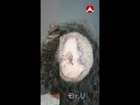
Dr. Umar, AKA Dr. Bumpinator, utilizes a meticulous excision procedure for FD lesions with minimal tissue damage. He also uses Guarded High-Tension Sutures to guide wound contraction, ensuring optimal healing and reducing the risk of recurrence.

First image represents the patient’s vertex immediately after surgical excision. Second image shows suture guards used to secure the operated area to prevent tissue strangulation. Third image is post-operation. Notice the use of high-tension suture guards.

Skin Grafting

After lesion excision, wounds that do not close and exceed a certain diameter length due to the surgical process will require skin grafting.

Below are Real-Life Patient Photos and Videos 

Preoperative FD plaque involving the vertex, mid-scalp extending to the frontal scalp. (A) Thirteen months after complete excision of FD lesion and healing by second intention, aided by high-tension sutures with guards and minor skin graft (B).

Frame A shows the patient after an operation and application of guarded high-tension sutures. Frame B is eight weeks post-operation. The doctor used a split-thickness skin graft from the inner thigh to close the open wound.

Frame A represents Folliculitis Decalvans covering the entire vertex before excision. Frame B is the same patient eight months after operation and second-intention healing. The doctor used high-tension sutures and skin grafts for the above patient.

Frame A represents Folliculitis Decalvans on the right parietal prominence area before surgical excision. Frame B is the same patient 9 months after lesion excision and second-intention healing with high-tension sutures.

Preoperative FD plaque involving the vertex, mid-scalp extending to the frontal scalp, and acne keloidalis nuchae in the nape area (A) and nineteen months after complete excision of FD lesion and healing by second intention, aided by guarded high-tension sutures and a minor STSG (Green arrow). The tenting skin has flattened out (yellow arrows (B).

FD plaque involving the right parietal prominence area before surgical excision and AKN lesions in the nape zone. (A) and four months after complete excision of the FD lesion and healing by second intention, aided by guarded high-tension sutures and a minor skin graft (B).

VIDEO: Folliculitis Decalvans Surgical Excision Before and After


Patient Selection Criteria Developed by Dr. Bumpinator

Dr. Sanusi Umar, known for his innovative approach to treating Folliculitis Decalvans (FD), credits his success to careful patient diagnosis and selection of appropriate treatment methods.

FD presents with varying lesion sizes and affects different areas of the scalp, meaning that a single treatment approach may not be practical for all patients. Recognizing this, Dr. Umar has developed tailored treatment plans.

By customizing treatment according to the extent and location of FD lesions, Dr. Umar ensures that each patient receives the most effective care, giving better results. 

An illustration of hair ‘tufting’ and crusting scalp on a patient with severe Folliculitis Decalvans (FD).

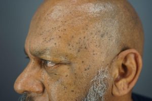
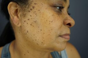
What Does Folliculitis Decalvans Look Like?

FD occurs as a result of inflamed hair follicles. Trapped bacteria causes acne-like red and pus-filled lesions on the affected areas, leading to pain and itchiness.

Who Gets Folliculitis Decalvans?

Folliculitis Decalvans can affect both men and women. In addition, there are higher incidences of FD in people of color. Learn more about Folliculitis Decalvans here. 

If you are struggling with Folliculitis Decalvans and want to speak to Dr.Bumpinator, click the free consultation button below to get started.


                                                                            FREE CONSULTATION  

References

Umar, Sanusi, et al. “Refractory Folliculitis Decalvans Treatment Success with a Novel Surgical Excision Approach Using Guarded High-Tension Sutures.” Clinical, Cosmetic and Investigational Dermatology, vol. Volume 16, 1 Sept. 2023, pp. 2381–2390, https://doi.org/10.2147/ccid.s422077.

Also Read

https://bumpinator.com/ear-keloid-treatment-that-youll-be-happy-with-permanently/

https://bumpinator.com/bumpinator/what-is-folliculitis-decalvans/


What is a Skin Tag or Dermatosis Papulosa Nigra (DPN)?

Dermatosis Papulosa Nigra (DPN), often referred to as skin tags, is a common dermatological condition that presents as small, benign lesions on the skin. While generally harmless, these dark, raised papules can cause cosmetic concerns for many individuals, affecting their self-esteem. Understanding DPN’s symptoms, causes, diagnosis, and treatment options can empower those affected to effectively manage and address this condition, as knowledge is key to power.

What is Dermatosis Papulosa Nigra (DPN)?

Dermatosis Papulosa Nigra (DPN) is a common, benign skin condition characterized by small, dark, raised lesions on the skin. Although it primarily affects individuals with darker skin tones, it can occur in all skin types. These lesions, known as skin tags, are generally harmless but can be cosmetically concerning as more and more appear.

DPN can be easy to spot as as skin tags develop, such as on the skin of this patient – notice the raised, dark bumps present.

DPN as a Variation of Seborrheic Keratosis

Dermatosis Papulosa Nigra (DPN) is considered by some experts to be a variation of seborrheic keratosis. Histologically, DPN resembles seborrheic keratoses and shares a mutation in FGFR3 with seborrheic keratoses. However, it does not involve a mutation in PIK3CA. The condition may be cosmetically undesirable to some patients.

Symptoms of Dermatosis Papulosa Nigra (DPN)

Dermatosis Papulosa Nigra DPN presents with distinctive features that can help in its identification even from home:

Primary Symptoms

  • Small, Dark Papules: These are the hallmark of DPN, appearing as dark brown or black papules ranging from 1-5 mm in diameter. They are flexible to the touch and easy to measure from side to side.
  • Raised Lesions: The papules are typically raised and can have a smooth or rough surface.
  • Location: Commonly found on the face, particularly around the cheeks and eyes, but can also appear on the neck, chest, and back.

Secondary Symptoms

  • Cosmetic Concerns: The appearance of these lesions can be considered cosmetically unattractive.
  • Itching or Irritation: In some cases, the papules may become infected or irritated if rubbed or scratched repeatedly on purpose or by mistake, such as by clothing rubbing the papules during everyday movement.

What Causes Dermatosis Papulosa Nigra (DPN)?

The exact cause remains unclear, but several factors are believed to contribute to DPN’s development:

  1. Genetics:
    There is a strong genetic component to DPN, as it often runs in families. Individuals with a family history of DPN are more likely to develop the condition.
  2. Age:
    DPN is commonly found in middle-aged and older adults. However, DPNs can be seen in patients of any age.
  3. Skin Type:
    DPN predominantly affects individuals with darker skin tones, particularly those of African descent.
  4. Nevoid Developmental Defects: Dermatosis Papulosa Nigra is can be caused by a nevoid developmental defect of the pilosebaceous follicle.

Epidemiology and Demographics

A study in the International Journal of Dermatology states that DPN tends to occur in females with Fitzpatrick skin types IV to VI. The lesions commonly develop in the head and neck. They are often associated with a family history of DPN and significant sun exposure.

Like the other patient above, notice the dark, raised spots on her skin – the telltale sign of DPN, many skin tags.

How is DPN Diagnosed?

Diagnosis of DPN is typically straightforward and involves:

  1. Physical Examination:
    Dermatologists can diagnose DPN by visually examining the skin and identifying the characteristic papules to clear out other causes or skin disorders like cancers.
  2. Dermatoscopy:
    In some cases, dermatoscopy may examine the lesions more closely to differentiate them from other skin conditions.
  3. Biopsy:
    Rarely, a skin biopsy may be ordered to rule out other conditions.

Severity Classification

Though benign, dermatologists classify DPN lesions according to severity:

  • Fewer than ten papules: Mild
  • More than ten papules: Moderate
  • Greater than fifty papules: Severe

DPN vs. Regular Moles

DPN is unlike regular skin moles, which are more aligned with skin follicles. Moles occur when skin cells, known as melanocytes, clump up. Melanocytes produce melanin, which colors our skin. While normally evenly distributed, they will occasionally come together. A malignant growth tends to be an asymmetrical mole. DPN lesions, where one side is a different shape from the other, are pretty standard. DPN bumps both increase in size and grow in number over time.

How Do You Treat Dermatosis Papulosa Nigra (DPN)?

Treatment of DPN is generally not medically necessary but may be sought for cosmetic reasons.

Cosmetic Treatments

  • Cryotherapy: Freezing the papules with liquid nitrogen to remove them.
  • Electrosurgery: Using electrical currents to remove the papules.
  • Laser Therapy: Utilizing ablative laser technology to target and remove the lesions precisely.
  • Excision: Surgically cutting out the papules, usually performed by a dermatologist.

Topical Treatments

  • Retinoids: Topical retinoid creams can sometimes help reduce the appearance of DPN, although their effectiveness varies.

Self-Care

  • Avoid Irritation: Patients are advised to avoid scratching or rubbing the lesions to prevent irritation.
  • Regular Monitoring: Regular check-ups with a dermatologist to monitor the condition and manage any changes.

Frequently Asked Questions (FAQ)

  • Is Dermatosis Papulosa Nigra treatable? The condition benign and doesn’t call for treatment unless it affects a person’s lifestyle; several cosmetic procedures can effectively remove the lesions or reduce their appearance.
  • Is Dermatosis Papulosa Nigra hereditary? Yes, there is a significant genetic component, making it more common in individuals with a family history of DPN.
  • What causes Dermatosis Papulosa Nigra? The exact cause is unknown, but genetics, age, and skin type are contributing factors. Practicing sun safety and having a skincare routine are extraordinary preventative measures.
  • How can I treat Dermatosis Papulosa Nigra? Treatment options include cryotherapy, electrosurgery, laser therapy, and excision. Consult a dermatologist for the best approach. Complete a free consultation for your hair loss with board-certified dermatologist Dr. Sanusi Umar to see how the real-life Bumpinator can help you!
  • Can Dermatosis Papulosa Nigra be prevented? Since the exact cause of DPN is not fully understood and it often runs in families, prevention is not clearly defined outside of making smart, generally skin-safe decisions.

References

Furukawa, Fumina et al. “Treatment of dermatosis papulosa nigra using a carbon dioxide laser.” Journal of cosmetic dermatology vol. 19,10 (2020): 2572-2575. doi:10.1111/jocd.13309

Maghfour, Jalal, and Temitayo Ogunleye. “A Systematic Review on the Treatment of Dermatosis Papulosa Nigra.” Journal of drugs in dermatology : JDD vol. 20,4 (2021): 467-472. doi:10.36849/JDD.2021.5555

Nowfar-Rad, Mehran, MD; Elston, Dirk M., MD. “Dermatosis Papulosa Nigra.” Medscape, Jun 07, 2022.

Tran, Mimi, and Vincent Richer. “Elective Treatment of Dermatosis Papulosa Nigra: A Review of Treatment Modalities.” Skin therapy letter vol. 25,4 (2020): 1-5.

Xiao, Anny, et al. “Dermatosis Papulosa Nigra.” StatPearls, StatPearls Publishing, 7 August 2023.