Folliculitis Decalvans Treatment

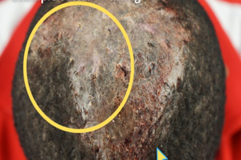
Do You Want to Get a Permanent Solution to Your Folliculitis Decalvans (FD) Condition? 

Folliculitis Decalvans (FD) is a form of scarring Alopecia that is difficult to treat and manage. Current treatment options used to manage FD often offer temporary solutions. 

A Smarter and Better Surgery for Folliculitis Decalvans

There is a way to permanently eliminate folliculitis decalvans. Four men and a woman suffering from the condition received treatment by surgically removing lesions from the affected areas.

The methods include: 

  •         Surgical excision
  •         Second-intention healing

Surgical Excision and Second-Intention Healing

A recent Clinical Cosmetic and Investigative Dermatology study described a novel procedure for treating Folliculitis Decalvans. This technique involved a specialized method of excising the lesions and applying patented Athena Suture Kits to facilitate wound closure. The authors observed that this method effectively and permanently eliminated Folliculitis Decalvans. Notably, the approach successfully employed second-intention wound healing on typically convex surfaces, a departure from its usual application on concave terrains.

Success Stories Published in a Medical Peer-Reviewed Study

Dr. Sanusi Umar, a pioneer in the field, became the first to perform the surgical excision of Folliculitis Decalvans (FD), a chronic and challenging scalp condition. This groundbreaking procedure was highlighted in a peer-reviewed medical study where five patients with varying severities of FD underwent surgery to remove their FD lesions.

According to the Clinical Cosmetic and Investigative Dermatology publication, these patients had previously tried other treatment methods without success. The thickness of their FD lesions made them unsuitable candidates for alternative treatments like laser therapy, radiation, and other conventional approaches.

The medical publication also highlighted that within 18 months, Dr. Umar successfully performed all five surgeries at his Los Angeles clinic, permanently eliminating the FD condition.

Dr. Umar, AKA Dr. Bumpinator, utilizes a meticulous excision procedure for FD lesions with minimal tissue damage. He also uses Guarded High-Tension Sutures to guide wound contraction, ensuring optimal healing and reducing the risk of recurrence.

First image represents the patient’s vertex immediately after surgical excision. Second image shows suture guards used to secure the operated area to prevent tissue strangulation. Third image is post-operation. Notice the use of high-tension suture guards.

Skin Grafting

After lesion excision, wounds that do not close and exceed a certain diameter length due to the surgical process will require skin grafting.

Below are Real-Life Patient Photos and Videos 

Preoperative FD plaque involving the vertex, mid-scalp extending to the frontal scalp. (A) Thirteen months after complete excision of FD lesion and healing by second intention, aided by high-tension sutures with guards and minor skin graft (B).

Frame A shows the patient after an operation and application of guarded high-tension sutures. Frame B is eight weeks post-operation. The doctor used a split-thickness skin graft from the inner thigh to close the open wound.

Frame A represents Folliculitis Decalvans covering the entire vertex before excision. Frame B is the same patient eight months after operation and second-intention healing. The doctor used high-tension sutures and skin grafts for the above patient.

Frame A represents Folliculitis Decalvans on the right parietal prominence area before surgical excision. Frame B is the same patient 9 months after lesion excision and second-intention healing with high-tension sutures.

Preoperative FD plaque involving the vertex, mid-scalp extending to the frontal scalp, and acne keloidalis nuchae in the nape area (A) and nineteen months after complete excision of FD lesion and healing by second intention, aided by guarded high-tension sutures and a minor STSG (Green arrow). The tenting skin has flattened out (yellow arrows (B).

FD plaque involving the right parietal prominence area before surgical excision and AKN lesions in the nape zone. (A) and four months after complete excision of the FD lesion and healing by second intention, aided by guarded high-tension sutures and a minor skin graft (B).

VIDEO: Folliculitis Decalvans Surgical Excision Before and After


Patient Selection Criteria Developed by Dr. Bumpinator

Dr. Sanusi Umar, known for his innovative approach to treating Folliculitis Decalvans (FD), credits his success to careful patient diagnosis and selection of appropriate treatment methods.

FD presents with varying lesion sizes and affects different areas of the scalp, meaning that a single treatment approach may not be practical for all patients. Recognizing this, Dr. Umar has developed tailored treatment plans.

By customizing treatment according to the extent and location of FD lesions, Dr. Umar ensures that each patient receives the most effective care, giving better results. 

An illustration of hair ‘tufting’ and crusting scalp on a patient with severe Folliculitis Decalvans (FD).

What Does Folliculitis Decalvans Look Like?

FD occurs as a result of inflamed hair follicles. Trapped bacteria causes acne-like red and pus-filled lesions on the affected areas, leading to pain and itchiness.

Who Gets Folliculitis Decalvans?

Folliculitis Decalvans can affect both men and women. In addition, there are higher incidences of FD in people of color. Learn more about Folliculitis Decalvans here. 

If you are struggling with Folliculitis Decalvans and want to speak to Dr.Bumpinator, click the free consultation button below to get started.


                                                                            FREE CONSULTATION  

References

Umar, Sanusi, et al. “Refractory Folliculitis Decalvans Treatment Success with a Novel Surgical Excision Approach Using Guarded High-Tension Sutures.” Clinical, Cosmetic and Investigational Dermatology, vol. Volume 16, 1 Sept. 2023, pp. 2381–2390, https://doi.org/10.2147/ccid.s422077.

Also Read

https://bumpinator.com/ear-keloid-treatment-that-youll-be-happy-with-permanently/

https://bumpinator.com/bumpinator/what-is-folliculitis-decalvans/


Dissecting Cellulitis Treatment

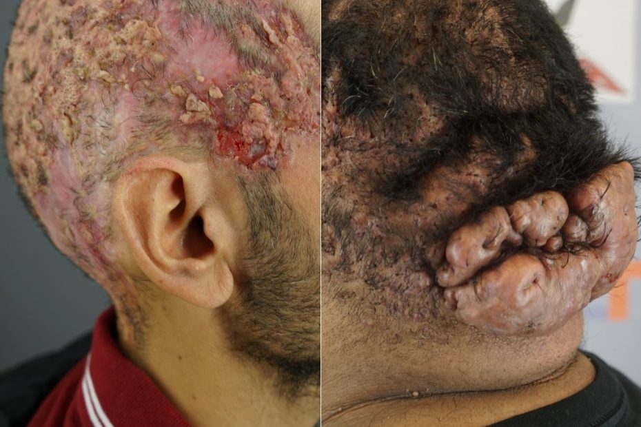
Dissecting Cellulitis (DC) is a long-term cicatricial (scarring) form of alopecia (hair loss) characterized by abscesses and keloids on the infected areas.

This condition has no cure; however, dermatologists recommend various treatment methods to manage the keloids and pus-filled lesions that occur as the condition progresses.

In rare cases, patients with severe scalp problems may simultaneously exhibit DC, Acne Keloidalis Nuchae (AKN), and Cutis Verticis Gyrata (CVG) symptoms.

A patient exhibiting Dissecting Cellulitis (DC), Acne Keloidalis Nuchae (AKN), and Cutis Verticis Gyrata (CVG).

Dr. Sanusi Umar, AKA The Bumpinator, devised a way to permanently eliminate the DC condition, which is particularly suitable for patients with other underlying conditions.

A Smarter and Better Surgery Treatment for DC with the Bumpinator

There are three ways to treat DC:

  •         Surgical Treatment

Surgically removing DC lesions from an infected scalp can leave extensive scarring. Dr. Umar devised an innovative and less invasive way to permanently treat DC through surgical excisions and uniquely closing up the wounds to acquire quality esthetic results.

Dr. Bumpinator’s methods are thorough to prevent reoccurrence and leave minimal scarring after treatment. Using his patented Athena Suture Kits, The Bumpinator successfully eliminates DC permanently.

  •         Laser Removal

Dr. Bumpinator employs the use of laser therapy to treat DC to reduce the side effects that result from prolonged use of medications and systemic treatments. 

  •         Radiation Therapy

In cases where patients have severe DC, Dr. Umar uses radiation therapy.

Dr. Sanusi Umar, aka Dr. Bumpinator, uses these as well as many innovative treatments he developed himself to accomplish the goal of long-term relief for DC patients with minimal side effects and footprints. Patients travel from all corners of the globe to receive treatment under Dr. Bumpinator’s care, following stories like the one in the case of Kevin.

The case highlighted in the video below shows a severe Dissecting Cellulitis covering the entire scalp.


Why is it Hard to Treat Dissecting Cellulitis (DC) Condition Permanently?

Current medications and topical steroids used in the treatment of DC provide temporary solutions. Patients have ended up in Dr. Umar’s clinic after failed attempts to remove the condition.

A common observation among patients under the Bumpinator’s care is the misdiagnosis of DC and AKN as razor bumps or a form of dermatitis, which leads to prolonged treatment with poor results.

Patient Selection Criteria Developed by Dr. Bumpinator

Some patients exhibit severe DC that require unique treatment methods, tailor-made as advised by a specialist after a careful diagnosis.

The patient depicted in the images below has a severe case of DC. He has been receiving treatment under Dr.Umar’s care to manage the condition using surgical treatment, radiation, and laser treatment.

This patient underwent treatment from different doctors, from monthly injections to unending follow-up visits to the hospital, and the condition worsened with time instead of improving.

A patient with a very severe case of dissecting cellulitis. He is currently receiving laser treatment under the care of Dr. Umar to manage symptoms.

If you notice signs that resemble those of Dissecting Cellulitis (DC), it is advisable to consult a certified board dermatologist like Dr. Sanusi Umar. Learn more about Dissecting Cellulitis (DC) here. 


To book a free consultation with Dr. Sanusi Umar, AKA Dr. Bumpinator, click the button below. 

                                                                            FREE CONSULTATION  

References

KRASNER, BRETT D., et al. “Dissecting Cellulitis Treated with the Long-Pulsed Nd:YAG Laser.” Dermatologic Surgery, vol. 32, no. 8, Aug. 2006, pp. 1039–1044, https://doi.org/10.1111/j.1524-4725.2006.32227.x. Accessed 15 Feb. 2021.

Umar, Sanusi, et al. “Refractory Folliculitis Decalvans Treatment Success with a Novel Surgical Excision Approach Using Guarded High-Tension Sutures.” Clinical, Cosmetic and Investigational Dermatology, vol. Volume 16, 1 Sept. 2023, pp. 2381–2390, https://doi.org/10.2147/ccid.s422077.

Also Read

https://bumpinator.com/ear-keloid-treatment-that-youll-be-happy-with-permanently/

https://bumpinator.com/bumpinator/what-is-folliculitis-decalvans/