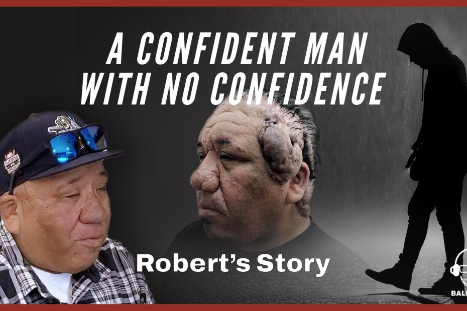
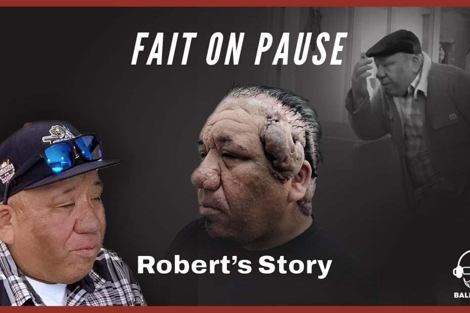
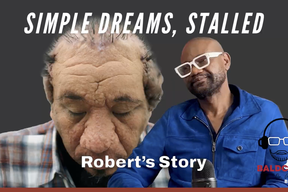
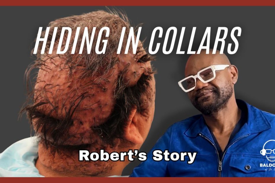
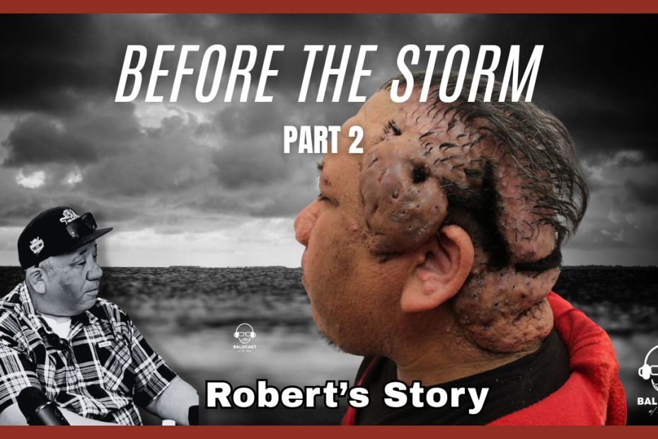
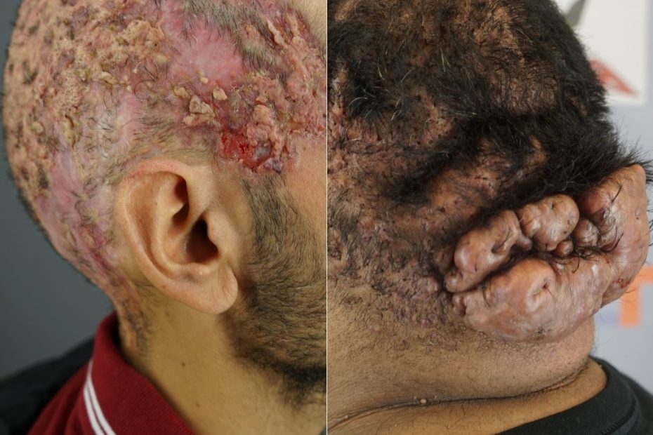
What is a Skin Tag or Dermatosis Papulosa Nigra (DPN)?

Dermatosis Papulosa Nigra (DPN), often referred to as skin tags, is a common dermatological condition that presents as small, benign lesions on the skin. While generally harmless, these dark, raised papules can cause cosmetic concerns for many individuals, affecting their self-esteem. Understanding DPN’s symptoms, causes, diagnosis, and treatment options can empower those affected to effectively manage and address this condition, as knowledge is key to power.

What is Dermatosis Papulosa Nigra (DPN)?

Dermatosis Papulosa Nigra (DPN) is a common, benign skin condition characterized by small, dark, raised lesions on the skin. Although it primarily affects individuals with darker skin tones, it can occur in all skin types. These lesions, known as skin tags, are generally harmless but can be cosmetically concerning as more and more appear.

DPN can be easy to spot as as skin tags develop, such as on the skin of this patient – notice the raised, dark bumps present.

DPN as a Variation of Seborrheic Keratosis

Dermatosis Papulosa Nigra (DPN) is considered by some experts to be a variation of seborrheic keratosis. Histologically, DPN resembles seborrheic keratoses and shares a mutation in FGFR3 with seborrheic keratoses. However, it does not involve a mutation in PIK3CA. The condition may be cosmetically undesirable to some patients.

Symptoms of Dermatosis Papulosa Nigra (DPN)

Dermatosis Papulosa Nigra DPN presents with distinctive features that can help in its identification even from home:

Primary Symptoms

  • Small, Dark Papules: These are the hallmark of DPN, appearing as dark brown or black papules ranging from 1-5 mm in diameter. They are flexible to the touch and easy to measure from side to side.
  • Raised Lesions: The papules are typically raised and can have a smooth or rough surface.
  • Location: Commonly found on the face, particularly around the cheeks and eyes, but can also appear on the neck, chest, and back.

Secondary Symptoms

  • Cosmetic Concerns: The appearance of these lesions can be considered cosmetically unattractive.
  • Itching or Irritation: In some cases, the papules may become infected or irritated if rubbed or scratched repeatedly on purpose or by mistake, such as by clothing rubbing the papules during everyday movement.

What Causes Dermatosis Papulosa Nigra (DPN)?

The exact cause remains unclear, but several factors are believed to contribute to DPN’s development:

  1. Genetics:
    There is a strong genetic component to DPN, as it often runs in families. Individuals with a family history of DPN are more likely to develop the condition.
  2. Age:
    DPN is commonly found in middle-aged and older adults. However, DPNs can be seen in patients of any age.
  3. Skin Type:
    DPN predominantly affects individuals with darker skin tones, particularly those of African descent.
  4. Nevoid Developmental Defects: Dermatosis Papulosa Nigra is can be caused by a nevoid developmental defect of the pilosebaceous follicle.

Epidemiology and Demographics

A study in the International Journal of Dermatology states that DPN tends to occur in females with Fitzpatrick skin types IV to VI. The lesions commonly develop in the head and neck. They are often associated with a family history of DPN and significant sun exposure.

Like the other patient above, notice the dark, raised spots on her skin – the telltale sign of DPN, many skin tags.

How is DPN Diagnosed?

Diagnosis of DPN is typically straightforward and involves:

  1. Physical Examination:
    Dermatologists can diagnose DPN by visually examining the skin and identifying the characteristic papules to clear out other causes or skin disorders like cancers.
  2. Dermatoscopy:
    In some cases, dermatoscopy may examine the lesions more closely to differentiate them from other skin conditions.
  3. Biopsy:
    Rarely, a skin biopsy may be ordered to rule out other conditions.

Severity Classification

Though benign, dermatologists classify DPN lesions according to severity:

  • Fewer than ten papules: Mild
  • More than ten papules: Moderate
  • Greater than fifty papules: Severe

DPN vs. Regular Moles

DPN is unlike regular skin moles, which are more aligned with skin follicles. Moles occur when skin cells, known as melanocytes, clump up. Melanocytes produce melanin, which colors our skin. While normally evenly distributed, they will occasionally come together. A malignant growth tends to be an asymmetrical mole. DPN lesions, where one side is a different shape from the other, are pretty standard. DPN bumps both increase in size and grow in number over time.

How Do You Treat Dermatosis Papulosa Nigra (DPN)?

Treatment of DPN is generally not medically necessary but may be sought for cosmetic reasons.

Cosmetic Treatments

  • Cryotherapy: Freezing the papules with liquid nitrogen to remove them.
  • Electrosurgery: Using electrical currents to remove the papules.
  • Laser Therapy: Utilizing ablative laser technology to target and remove the lesions precisely.
  • Excision: Surgically cutting out the papules, usually performed by a dermatologist.

Topical Treatments

  • Retinoids: Topical retinoid creams can sometimes help reduce the appearance of DPN, although their effectiveness varies.

Self-Care

  • Avoid Irritation: Patients are advised to avoid scratching or rubbing the lesions to prevent irritation.
  • Regular Monitoring: Regular check-ups with a dermatologist to monitor the condition and manage any changes.

Frequently Asked Questions (FAQ)

  • Is Dermatosis Papulosa Nigra treatable? The condition benign and doesn’t call for treatment unless it affects a person’s lifestyle; several cosmetic procedures can effectively remove the lesions or reduce their appearance.
  • Is Dermatosis Papulosa Nigra hereditary? Yes, there is a significant genetic component, making it more common in individuals with a family history of DPN.
  • What causes Dermatosis Papulosa Nigra? The exact cause is unknown, but genetics, age, and skin type are contributing factors. Practicing sun safety and having a skincare routine are extraordinary preventative measures.
  • How can I treat Dermatosis Papulosa Nigra? Treatment options include cryotherapy, electrosurgery, laser therapy, and excision. Consult a dermatologist for the best approach. Complete a free consultation for your hair loss with board-certified dermatologist Dr. Sanusi Umar to see how the real-life Bumpinator can help you!
  • Can Dermatosis Papulosa Nigra be prevented? Since the exact cause of DPN is not fully understood and it often runs in families, prevention is not clearly defined outside of making smart, generally skin-safe decisions.

References

Furukawa, Fumina et al. “Treatment of dermatosis papulosa nigra using a carbon dioxide laser.” Journal of cosmetic dermatology vol. 19,10 (2020): 2572-2575. doi:10.1111/jocd.13309

Maghfour, Jalal, and Temitayo Ogunleye. “A Systematic Review on the Treatment of Dermatosis Papulosa Nigra.” Journal of drugs in dermatology : JDD vol. 20,4 (2021): 467-472. doi:10.36849/JDD.2021.5555

Nowfar-Rad, Mehran, MD; Elston, Dirk M., MD. “Dermatosis Papulosa Nigra.” Medscape, Jun 07, 2022.

Tran, Mimi, and Vincent Richer. “Elective Treatment of Dermatosis Papulosa Nigra: A Review of Treatment Modalities.” Skin therapy letter vol. 25,4 (2020): 1-5.

Xiao, Anny, et al. “Dermatosis Papulosa Nigra.” StatPearls, StatPearls Publishing, 7 August 2023.

What is PIILIF?

What is PIILIF (Perifollicular Infundibulo-Isthmic Lymphocytoplasmic Infiltrates and Fibrosis)? It is a dermatological condition characterized by inflammation of hair follicles, hair loss, and scalp scarring. Recent research suggests that the condition affects scalp hair and other non-scalp hair follicles. In rare cases, PIILIF might involve the eyebrows, eyelashes, beards, and pubic regions.

Perifollicular Infundibulo-Isthmic Lymphocytoplasmic Infiltrates and Fibrosis is often ignored and mistaken for seborrheic dermatitis or dandruff, especially in cases where patients fail to manifest clinical symptoms.

What Causes PIILIF?

Researchers have not yet established the cause of Perifollicular Infundibulo-Isthmic Lymphocytoplasmic Infiltrates and Fibrosis. It likely signifies a low-level autoimmune attack of the hair. It is likely the precursor to currently recognized autoimmune scarring alopecias such as lichen planopilaris – LPP, frontal fibrosing alopecia – FFA, central centrifugal cicatricial alopecia – CCCA, acne keloidalis nuchae – AKN, Folliculitis decalvans – FD, dissecting cellulitis – DC. PIILIF  is largely influenced by several factors, such as; 

  • Genetics.
  • Hormonal. For instance, AKN and DC overwhelmingly affect men at the onset of puberty.
  • Environmental Triggers. Exposure to chemicals, allergens, pollutants, trauma, or scalp injuries. 
  • Scalp location
  • Infection/superantigen

Contexts Where Can PIILIF Be Found

Primary Condition

A doctor can diagnose PIILIF as a standalone condition with histopathological features and symptoms such as fibrosis, inflammation, and hair loss. However, it does not necessarily indicate the presence of other underlying conditions.

PIILIF Shared Precursor Condition for Acne Keloidalis Nuchae (AKN) and Primary Cicatricial Alopecias (PCAs)

Understanding the relationship between PIILIF and other Primary Cicatricial Alopecias (PCAs) can help diagnose and manage the conditions early to prevent progression.

A research study by Dr. Sanusi Umar, conducted on 41 male patients suffering from AKN, showed symptoms and signs of PIILIF. Moreover, the signs were also exhibited on the Normal-appearing scalp (NAS), away from the AKN lesions. The findings further highlighted the presence of scarring, inflammation, and scaling on some of the patients’ beards and other hairy body parts. This was evidence that PIILIF is a scalp condition and affects hair follicles in different body parts.

An illustration of PIILIF as a shared precursor condition to Primary Cicatricial Alopecias (PCAs).

Yet, other studies have established the occurrence of PIILIF in the normal-appearance scalp zones in the following classical scarring alopecia: LPP, CCCA, FFA, AKN, FD, and Dissecting cellulitis.

PIILIF(Perifollicular Infundibulo-Isthmic Lymphocytoplasmic Infiltrates and Fibrosis) Symptoms

  •         No symptoms
  •         +/- Hair loss  (Alopecia)
  •          +/- Itching, redness, and inflammation of the scalp
  •          +/- Scaliness/flaking

In addition, other symptoms may include early stages of Primary Cicatricial Alopecias (PCAs).


How Do You Diagnose PIILIF?

A clinical diagnosis is recommended for PIILIF, as the symptoms may overlap with those of other underlying conditions.

PIILIF diagnosis methods include:

  • Physical examination. A specialist will monitor visible symptoms on the scalp and other hairy body parts, such as the beard, eyebrows, and eyelashes.
  • Trichoscopy. This method evaluates skin lesions on the affected areas under a magnifying lens and lighting to confirm the presence of PIILIF.
  • Biopsy: A sample of hair or scalp skin is observed under a microscope to ascertain the PIILIF condition before prescribing further treatment.

How to Treat PIILIF(Perifollicular Infundibulo-Isthmic Lymphocytoplasmic Infiltrates and Fibrosis)

Although PILLIF cannot be cured, health practitioners have devised several treatment options to manage the condition and prevent its progression.

Another key point to note is that treating Perifollicular Infundibulo-Isthmic Lymphocytoplasmic Infiltrates and Fibrosis requires addressing other possible underlying conditions to counter the diseases in their early stages. This is because underlying conditions more often lead to severe PIILIF. 

Treatment options include:

  • Oral Medications:  Tetracyclines for their anti-inflammatory properties.
  • Topical Treatments. Consider Tacrolimus vs Pemicrolimin. 
  • Oral Supplements: EG Gashee oral.

PIILIF(Perifollicular Infundibulo-Isthmic Lymphocytoplasmic Infiltrates and Fibrosis) Frequently Asked Questions (FAQ)

Can PIILIF be cured?

There is no cure; however, an early diagnosis can prevent progression, and various treatment methods can manage the symptoms. 

Should I see a specialist for PIILIF?

Yes. Consult a certified board dermatologist for a proper diagnosis, as it might be a precursor for other diseases.

Finally, you can get a free consultation with Dr. Sanusi Umar through this link: https://dru.com/hair-free-consultation/ or by using the consultation button below. 

References  

Doche, Isabella, et al. “Acne Keloidalis Nuchae and Folliculitis Decalvans: Same Process Affecting the Follicle or Coexisting Diseases? A Retrospective Study.” International Journal of Dermatology, vol. 58, no. 10, 26 June 2019, https://pubmed.ncbi.nlm.nih.gov/31241169/

Umar, Sanusi, et al. A Retrospective Cohort Study and Clinical Classification System of Acne Keloidalis Nuchae. Vol. 14, no. 4, 1 Apr. 2021. Accessed 17 June 2024.

Umar, Sanusi, et al. “Unveiling a Shared Precursor Condition for Acne Keloidalis Nuchae and Primary Cicatricial Alopecias.” Clinical, Cosmetic and Investigational Dermatology, vol. Volume 16, 1 Aug. 2023, pp. 2315–2327, https://doi.org/10.2147/ccid.s422310. Accessed 17 June 2024.

Wang, Eddy H C, et al. “Primary Cicatricial Alopecias Are Characterized by Dysregulation of Shared Gene Expression Pathways.” PNAS Nexus, vol. 1, no. 3, 11 July 2022, https://doi.org/10.1093/pnasnexus/pgac111.

Dissecting Cellulitis Treatment

Dissecting Cellulitis (DC) is a long-term cicatricial (scarring) form of alopecia (hair loss) characterized by abscesses and keloids on the infected areas.

This condition has no cure; however, dermatologists recommend various treatment methods to manage the keloids and pus-filled lesions that occur as the condition progresses.

In rare cases, patients with severe scalp problems may simultaneously exhibit DC, Acne Keloidalis Nuchae (AKN), and Cutis Verticis Gyrata (CVG) symptoms.

A patient exhibiting Dissecting Cellulitis (DC), Acne Keloidalis Nuchae (AKN), and Cutis Verticis Gyrata (CVG).

Dr. Sanusi Umar, AKA The Bumpinator, devised a way to permanently eliminate the DC condition, which is particularly suitable for patients with other underlying conditions.

A Smarter and Better Surgery Treatment for DC with the Bumpinator

There are three ways to treat DC:

  •         Surgical Treatment

Surgically removing DC lesions from an infected scalp can leave extensive scarring. Dr. Umar devised an innovative and less invasive way to permanently treat DC through surgical excisions and uniquely closing up the wounds to acquire quality esthetic results.

Dr. Bumpinator’s methods are thorough to prevent reoccurrence and leave minimal scarring after treatment. Using his patented Athena Suture Kits, The Bumpinator successfully eliminates DC permanently.

  •         Laser Removal

Dr. Bumpinator employs the use of laser therapy to treat DC to reduce the side effects that result from prolonged use of medications and systemic treatments. 

  •         Radiation Therapy

In cases where patients have severe DC, Dr. Umar uses radiation therapy.

Dr. Sanusi Umar, aka Dr. Bumpinator, uses these as well as many innovative treatments he developed himself to accomplish the goal of long-term relief for DC patients with minimal side effects and footprints. Patients travel from all corners of the globe to receive treatment under Dr. Bumpinator’s care, following stories like the one in the case of Kevin.

The case highlighted in the video below shows a severe Dissecting Cellulitis covering the entire scalp.


Why is it Hard to Treat Dissecting Cellulitis (DC) Condition Permanently?

Current medications and topical steroids used in the treatment of DC provide temporary solutions. Patients have ended up in Dr. Umar’s clinic after failed attempts to remove the condition.

A common observation among patients under the Bumpinator’s care is the misdiagnosis of DC and AKN as razor bumps or a form of dermatitis, which leads to prolonged treatment with poor results.

Patient Selection Criteria Developed by Dr. Bumpinator

Some patients exhibit severe DC that require unique treatment methods, tailor-made as advised by a specialist after a careful diagnosis.

The patient depicted in the images below has a severe case of DC. He has been receiving treatment under Dr.Umar’s care to manage the condition using surgical treatment, radiation, and laser treatment.

This patient underwent treatment from different doctors, from monthly injections to unending follow-up visits to the hospital, and the condition worsened with time instead of improving.

A patient with a very severe case of dissecting cellulitis. He is currently receiving laser treatment under the care of Dr. Umar to manage symptoms.

If you notice signs that resemble those of Dissecting Cellulitis (DC), it is advisable to consult a certified board dermatologist like Dr. Sanusi Umar. Learn more about Dissecting Cellulitis (DC) here. 


To book a free consultation with Dr. Sanusi Umar, AKA Dr. Bumpinator, click the button below. 

                                                                            FREE CONSULTATION  

References

KRASNER, BRETT D., et al. “Dissecting Cellulitis Treated with the Long-Pulsed Nd:YAG Laser.” Dermatologic Surgery, vol. 32, no. 8, Aug. 2006, pp. 1039–1044, https://doi.org/10.1111/j.1524-4725.2006.32227.x. Accessed 15 Feb. 2021.

Umar, Sanusi, et al. “Refractory Folliculitis Decalvans Treatment Success with a Novel Surgical Excision Approach Using Guarded High-Tension Sutures.” Clinical, Cosmetic and Investigational Dermatology, vol. Volume 16, 1 Sept. 2023, pp. 2381–2390, https://doi.org/10.2147/ccid.s422077.

Also Read

https://bumpinator.com/ear-keloid-treatment-that-youll-be-happy-with-permanently/

https://bumpinator.com/bumpinator/what-is-folliculitis-decalvans/

What is Dissecting Cellulitis?

What is Dissecting Cellulitis? Dissecting Cellulitis (DC), also known as Hoffman disease, is a chronic (long-term) inflammatory scalp disorder that causes abscesses and keloids that lead to patchy hair loss and cicatricial (scarring) alopecia (destruction of hair follicles).

Dissecting cellulitis is incurable; however, somebody can manage symptoms to reduce pain and provide comfort.

DC is part of the follicular occlusion triad, where three diseases occur simultaneously. The other inflammatory diseases are acne conglobate (nodulocystic acne) and hidradenitis suppurativa (acne inversa).

The disease is most prominent in young adult black males but can, however, affect people of all races, ages, and sex.

What Causes Dissecting Cellulitis?

Specialists are working to determine the main cause of DC and establish why it affects some people and not others.

In this disorder, blocked hair follicles swell up and eventually rupture, causing inflammation underneath the skin.

Secondary Infection

The pus formed in the scalp following the follicle bursts becomes infected with bacteria over time, leading to further surface infections, hair loss, and scarring.

DC is often attributed as hereditary, but the disease rarely occurs in multiple family members. A small number of patients from the same family have experienced DC in the past, which leaves room for the assumption that genes play a role in the disease.  

Symptoms of Dissecting Cellulitis

DC, which starts as blocked hair follicles, worsens with time, with various signs and symptoms manifesting at the onset and later stages of the disease if not treated on time.

Early Symptoms

The most common onset symptoms include:

  • Swollen pimples on the scalp
  • Pus-filled painful lesions
  • Hair loss patches

Symptoms noticed in fewer cases:

  • Fever and swollen lymph nodes

Late Symptoms

  • Bleeding lesions
  • Permanent hair loss (fails to grow back after homecare treatments)
  • Wormlike skin fold between lesions
  • Scarring

If you exhibit the above symptoms, it is essential to seek immediate dermatology consultation, as they may indicate DC disease.

How is Dissecting Cellulitis Diagnosed?

Dermatologists usually diagnose DC through physical examinations of the scalp or infected areas. To further ascertain a DC diagnosis, they may swab pus (if the symptoms are at a later stage) or take scalp biopsies for histologic analysis.

Consult certified specialists and explain your symptoms in detail to avoid a misdiagnosis. 

What Does it Look Like?

A patient with a very severe case of dissecting cellulitis. He is currently receiving laser treatment under the care of Dr. Umar to manage symptoms.

What Treatments Are Available for Dissecting Cellulitis?

Once diagnosed, there are several recommended approaches to treating DC. As an incurable chronic condition, strategies have been put in place to manage active symptoms such as pain, hair loss, and lesions.

Medications 

There are three kinds of supplements used to manage DC:

  • Antibiotic oral supplements to fight infection. It is often recommended for long-term use to prevent relapse.
  • Steroid supplements are used in the short term to manage inflammation. They should be avoided in the long run to prevent severe side effects.
  • Oral retinoids help reduce follicle blockage and reduce acne. Retinoids cannot be purchased over the counter. Patients require a prescription from a certified dermatologist.

WARNING: Pregnant mothers are advised to refrain from retinoid use, which may cause extensive damage to the unborn child.

Topical Steroids

These are substances applied directly to the affected areas to reduce scalp inflammation. Examples include gels, lotions, ointments, and creams. These can be combined with topical antimicrobials and antiseptic scalp washes to reduce bacterial levels.

Surgery

Specialists may surgically drain large boils or painful abscesses to manage them by excising. Additionally, they can cut open the inflamed areas.

Surgical removal of the entire scalp skin is an extreme procedure and could be one of the most effective methods to manage DC. However, this procedure is for specific patients with severe cases. If the patient requests it, the doctor may follow other treatment methods for better results. 

Note: This exercise may leave scarring.

Other Treatments

  • Laser hair removal may lead to permanent hair loss.
  • Laser treatment to smoothen the scalp.
  • Radiation. 
  • Light treatment (photodynamic therapy). The procedure is slightly painful and might cause crusting. 
  • Steroid Injections are administered directly to affected areas. They are a painful procedure but have fewer side effects than steroid tablets.

Self-Care

  • Avoid smoking or tobacco use. Research findings have linked tobacco to cause DC.
  • Follow instructions and medications as directed.
  • Use medically approved products on your hair and scalp.
  • Maintain a normal, healthy diet.
  • Do regular check-ups with your specialist. 

A severe case of Dissecting Cellulitis (DC)

Dissecting Cellulitis (DC) and Acne Keloidalis Nuchae (AKN)

Dissecting Cellulitis can, in rare cases, coexist with other scalp and hair disorders.

Acne Keloidalis Nuchae, also known as AKN, is a skin disorder characterized by small or large bumps, often on the back of the head. Although common in males of African-American ethnicity, AKN can also affect people of other races.

AKN is often mistaken or misdiagnosed as razor bumps or keloidal conditions. It is far worse if left untreated. Use this link to ensure you get a diagnosis from a board-certified dermatologist like Dr. Sanusi Umar: https://dru.com/hair-free-consultation/

Some patients with severe scalp conditions may exhibit symptoms of both DC, AKN, and CVG (cutis verticis gyrata, a rare disorder common in males, characterized by deep skin folds on the scalp) due to overlapping risk factors. 

Additionally, multifaceted treatment is applied to ensure DC, AKN, and CVG are managed effectively. 

A patient with severe cutis verticis gyrata (CVG), acne keloidalis nuchae (AKN), and dissecting cellulitis (DC) conditions. He is currently receiving treatment under the care of Dr. Umar to manage symptoms. Notice the small and large bumps at the back of his head and the furrow-like appearance on the center of the scalp.


Dissecting Cellulitis Frequently Asked Questions (FAQ)

Is Dissecting Cellulitis Fatal?

Dissecting Cellulitis is not a deadly disease.

Is Dissecting Cellulitis Contagious?

No. Dissecting Cellulitis is not contagious and cannot be passed from one person to another.

Is Dissecting Cellulitis Curable?

No. It is incurable but can be managed.

How Can I Treat Dissecting Cellulitis?

You can complete a free consultation for your scalp condition with board-certified dermatologist Dr. Sanusi Umar using this link: https://dru.com/hair-free-consultation/ or the free consultation button below.

FREE CONSULTATION

References

Hintze, J. M., Howard, B. E., Donald, C. B., & Hayden, R. E. (2016). Surgical Management and Reconstruction of Hoffman’s Disease (Dissecting Cellulitis of the Scalp). Case Reports in Surgery, 2016, 1–4. https://doi.org/10.1155/2016/2123037

Tran, A. X., Lefante, J. J., & Murina, A. (2022). Risk factors for dissecting cellulitis of the scalp: A case-control study. Journal of the American Academy of Dermatology, 86(4), 941–943. https://doi.org/10.1016/j.jaad.2021.03.076

What is Cutis Verticis Gyrata (CVG) or Bulldog Scalp Syndrome?

What is cutis verticis gyrata (CVG)? CVG is a rare disorder common in males, characterized by deep skin folds on the scalp resembling those of the brain. Also known as the “bulldog scalp” syndrome, the disorder is classified into primary and secondary forms.

Primary Form

The primary form of bulldog scalp syndrome, or CVG, affects the vertex and occipital regions on the scalp, with folds running symmetrically in an anterior-posterior direction.

The two types are:

  •  Primary Essential CVG: This form is prevalent among young adults, especially postpubertal men. The cause is attributed to genetics and endocrine factors.
  •  Primary Non-essential CVG: Unlike the primary essential, the non-essential CVG is often associated with various abnormalities such as schizophrenia, epilepsy, retardation, ophthalmologic and cranial abnormalities.

Secondary Form

The secondary form of bulldog scalp syndrome, or CVG, is associated with inflammatory and underlying scalp diseases. These may include cutis laxa, syphilis, amyloidosis, intracranial aneurysms, and acromegaly. 

What Causes Cutis Verticis Gyrata (CVG)?

Little research on the disorder shows that the cause is still undetermined. Understanding the etiology of CVG shows that it is not an individual disease entry. It is a manifestation of diverse causes:

Hormonal: The post-puberty onset of CVG and the male dominance suggest that various hormones can cause the disorder.

Imbalances of the growth hormone (GH) in some cases may lead to acromegaly or other endocrine disorders, which affect the overall physical growth of the body.

Genetics:

Genetics is often linked to the primary non-essential CVG, where the form is associated with neurodevelopmental disorders like epilepsy and retardation, among other genetic syndromes such as Turner Syndrome and Acromegaly.

Chronic Inflammation: Persistent scalp infections with recurrent inflammations may affect the skin, developing CVG folds and ridges.

Symptoms of Cutis Verticis Gyrata (CVG)

Although CVG symptoms are distinctive and visible ridges and furrows, here are others to look out for:

Primary Symptoms

  • Ridges and Furrow: These are very noticeable from the onset, with soft folds varying in prominence and depth. The ridges and furrows grow gradually over time.
  • Thickened Skin: During the early stages, the skin on the scalp thickens and becomes sponge-like to the touch.

Secondary Symptoms

In the case of secondary CVG form, one might experience additional symptoms related to underlying disorders or conditions:

  •   Hair loss or thinning on the affected areas.
  •   Discomfort and itching
  •   Other skin and hair conditions
  •   Symptoms related to underlying disorders may include weight loss or gain, fatigue, and enlarged body parts. 

How is CVG Diagnosed?

Bulldog scalp syndrome is particularly easy and fast to diagnose. Dermatologists examine the scalp physically. The presence of furrows and ridges is sufficient to diagnose CVG.

In rare cases, a specialist will further assess other visible symptoms to classify them as primary or secondary CVG.

In the case of secondary CVG, diagnostic tests such as skin biopsy and blood tests may be ordered to check for underlying disorders such as thyroid, acromegaly, or other scalp inflammatory diseases.

Patients with CVG are advised to avoid self-treatment to ensure a correct diagnosis has been made by a certified dermatologist, who may, in turn, refer them to specialists based on the findings. 

What does Bulldog scalp syndrome look like? 

Two of our patients in the above images struggled with both Bulldog Scalp Syndrome/Cutis Verticis Gyrata (CVG) and Acne Keloidalis Nuchae (AKN). Notice the visible ridges and furrows on the scalp as well as the patchy hair loss.


How Do You Treat CVG?

It is important to determine the form of Bulldog scalp syndrome/CVG diagnosed (primary or secondary) before proceeding with the treatment to ensure the methods used are effective.

To properly treat CVG, specialists are required to manage the cosmetic needs associated with changes in the scalp skin and the underlying conditions leading to the effects on the skin.

Cosmetic Treatment

  1. Surgical procedures

The most effective treatment method is surgical intervention, which depends on the location and size of the CVG folds. Big folds may require skin grafts and local flap reconstructions, while smaller furrows can be managed with simple cuts out of the affected area.

Surgical procedures include:

  • Scalp reduction to flatten the surface.
  • Laser Therapy to make the scalp skin smooth.
  • Hair transplant
  1. Topical Treatments to manage dryness and itching.
  2. Use of corticosteroid injections to reduce inflammation.

Managing Underlying Conditions

  • Hormonal therapy
  • Treatment of associated dermatological conditions
  • Preventing progressive skin changes through essential treatment of underlying disorders.

Self-Care

  • Patients are encouraged to practice scalp hygiene by cleaning between furrows and ridges to prevent bad odors and secondary infections. Ensure products used are mild to the skin to reduce irritation.
  • Avoid tight headgear or hairstyles to minimize scalp trauma.

CVG patients, especially those with associated conditions, require regular monitoring by specialists to assess progress and address complications or adjust treatment.

Relationship Between CVG (Cutis Verticis Gyrata) and AKN (Acne Keloidalis Nuchae (AKN)

CVG and AKN are both scalp conditions prevalent in men and characterized by small and large folds on the back region of the head. A 2022 research study on the relationship between dermal thickening and excessive folding in both disorders revealed that the spreading of AKN lesions preceded the onset of CVG by two years for patients affected by the two disorders. 

In the wake of the research findings associating AKN and CVG, dermatologists have become more aware of the possibility of patients experiencing one after the other or both simultaneously. Specialists can now take precautions in addition to suggested treatment methods to manage the disorders. 

Frequently Cutis Verticis Gyrata Asked Questions (FAQ)

Is Cutis Verticis Gyrata treatable?

Treatment options include managing underlying conditions, symptoms, and cosmetic needs.

Is Cutis Verticis Gyrata hereditary?

Some forms of CVG contain genetic components. However, the disease is rare and patternless.

What causes Cutis Verticis Gyrata/Bulldog Scalp Syndrome? 

The exact cause is yet to be established. It can be either primary (spontaneous disease) or secondary (underlying conditions).

How can I treat Cutis Verticis Gyrata? 

Complete a free consultation for your hair loss with board-certified dermatologist Dr. Sanusi Umar by using this link: https://dru.com/hair-free-consultation/ or

Check out Dr. Bumpinator’s remarkable journeys with his patients using this link:  https://bumpinator.com/bumpinator/how-this-big-lucky-akn-bear-overcame-his-breaking-point-part-1/


References

Jeong, J., Daniela Tiemi Sano, Silvia Regina Martins, Antônio José Tebcherani, & Paula, A. (2014). Primary essential cutis verticis gyrata – Case report. Anais Brasileiros de Dermatologia, 89(2), 326–328. https://doi.org/10.1590/abd1806-4841.20142949

Koregol, S., Yatagiri, R., Warad, S., & Itagi, N. (2016). A rare association of scleromyxedema with cutis verticis gyrata. Indian Dermatology Online Journal, 7(3), 186. https://doi.org/10.4103/2229-5178.182365

Larsen, F., & Birchall, N. (2007). Cutis verticis gyrata: Three cases with different aetiologies that demonstrate the classification system. Australasian Journal of Dermatology, 48(2), 91–94. https://doi.org/10.1111/j.1440-0960.2007.00343.x


A New Beginning: Maria’s Liberation from 17 Years of Silent Suffering

In medicine, few stories encapsulate the essence of perseverance, resilience, and the transformative power of skilled medical intervention as profoundly as Maria’s journey under my care. Folliculitis Decalvans (FD) rarely affects women; Maria is one of the few who was dealt the unlucky card. But Maria’s story doesn’t stop there.

Today, I am honored to share this remarkable testament to the unwavering human spirit and the impact modern medicine and empathy can have on a patient’s life.

The Beginning of Maria’s Struggle

Maria’s health issues began innocently enough, with what appeared to be dandruff and flaking. Believing it to be a manageable problem, she turned to medicated shampoos to find relief. However, as her condition worsened and her scalp began oozing pus, Maria faced a difficult choice – she had to stop breastfeeding her newborn son due to the prescription of antibiotics.

FD in women: Maria’s scalp lesion kept growing and started oozing puss.

The Misdiagnosis

When the lesions on her scalp grew larger, Maria was told she had lupus and began a rigorous treatment plan involving strong medications. Two years later, she received the shocking revelation that she had been misdiagnosed. 

The news engulfed Maria in frustration and a sense of hopelessness. The prolonged journey of ineffective treatments had taken a toll on her physical and emotional well-being, intensifying the challenges she faced in regaining control of her health. 

Meanwhile, the bump on her head continued to grow, and the medications intended to help only weakened her immune system, leaving her physically drained.

Years of Searching for Answers

Maria endured 20 specialists and 17 years of uncertainty and suffering as she sought a correct diagnosis for her condition. The toll on her physical and emotional well-being was immeasurable. It affected not only her but also her children, who witnessed their mother in constant pain and discomfort. They found themselves taking on caretaker roles at a young age.

The Turning Point: The Discovery and Intervention

Finally, Maria’s journey led her to my clinic, where a thorough examination identified the bump on her head as a Folliculitis Decalvans (FD) lesion. Considering that FD seldom impacts women, Maria’s situation appeared exceptional. Treating unique cases poses greater challenges due to the scarcity of reference points.

Recognizing the gravity of her situation, I extended an invitation for surgery in my private clinic – an offer she graciously accepted, marking a pivotal moment in her path to recovery.

Athena to the Rescue 

To permanently remove the FD, I utilized my bat excision method, a surgical approach designed for optimal wound closure with secondary intention healing (SIH). Given the relatively larger height of Maria’s plaque, I employed my patented Athena sutures, designed specifically to assist with SIH.

The Athena suture guard kit (named after my beloved late dog Athena) assists with secondary intention healing after the FD surgery.

A New Lease on Life

A year post-surgery, Maria reflects on her remarkable transformation. Free from the burden of pain and discomfort, as her FD has not returned since the surgery, Maria is now leading a tumor and pain-free life.

“God has plans for us, and he put Dr. U in my path like an angel. He has given me a new chance at life, a life without pain”, she says.

Maria’s FD removal results in 11 months post-surgery. *

Maria’s Gratitude

Maria’s gratitude knows no bounds, as she acknowledges the surgery’s positive impact on her and her children’s lives. The indescribable relief she feels is a testament to the dedication and commitment of the healthcare profession, and I am humbled to have played a role in her journey to recovery.

Maria, pictured with Dr. Umar. She’s grateful to live FD-free now.

Conclusion

Maria’s story is a beacon of hope for those grappling with undiagnosed conditions. It underscores the importance of perseverance and the potential for transformative healing when a patient and a dedicated healthcare professional come together. I am delighted to have been part of Maria’s triumph over adversity, and her journey serves as a reminder of the profound impact we, as healthcare providers, can have on the lives of those under our care.

FREE CONSULTATION

Frequently Asked Questions

How can I know if I have FD?

If you suspect Folliculitis Decalvans (FD), consult a dermatologist for diagnosis and treatment. Common signs include firm bumps, itching, inflammation, and spots filled with pus may develop. 

FD predominantly affects men of African and Hispanic descent but can also occur in white men and rarely in women, as you saw in this article. 

Seek professional help for an accurate diagnosis and follow recommended treatments, such as medications or laser therapy, to manage FD effectively. Avoid self-diagnosis and treatment, as proper medical guidance is essential.

I was treated for FD, but now it’s back. What can I do?

Most treatments commonly practiced for Folliculitis Decalvans (FD) only offer a temporary solution and may lead to constant visits to the doctor over a long period.

Luckily, Dr. Sanusi Umar came up with a permanent solution.

Using his specialized procedures and inventions he created such as his patented Athena Suture Kits, Dr.Umar AKA The Bumpinator has been able to successfully eliminate Folliculitis Decalvans (FD) in several happy and satisfied

References

Lupus (no date) Mayo Clinic. Available at: https://www.mayoclinic.org/diseases-conditions/lupus/symptoms-causes/syc-20365789 (Accessed: 04 December 2023).

Umar, S. (2019) Is Dandruff – Seborrheic Dermatitis, A Cause of Hair Loss and Poor Hair Transplant Outcomes?, dru.com. Available at: https://dru.com/is-dandruff-seborrheic-dermatitis-a-cause-of-hair-loss-and-poor-hair-transplant-outcomes/ (Accessed: 04 December 2023). 

Umar, S. et al. (2019) Innovative Surgical Approaches and selection criteria of large acne Keloidalis nuchae lesions, Plastic and reconstructive surgery. Global open. Available at: https://www.ncbi.nlm.nih.gov/pmc/articles/PMC6571336/ (Accessed: 04 December 2023). 

Further Reading 

https://bumpinator.com/bumpinator/what-is-folliculitis-decalvans/

Multiple Large AKN Bumps Removal: AKN Surgery Success Before & After Results

AKN Removal Results Los Angeles: Another Successful Case by The Bumpinator

Understanding the Factors that Predispose You to Keloids

Keloids are raised scars that extend beyond the original wound, posing a mystery to researchers. Despite the unclear exact cause, various factors contribute to their development. This article delves into the elements that make someone prone to keloids, shedding light on the complex nature of this skin condition.

Genetics

Genetics significantly influences keloid development. Those with a family history or specific ethnic backgrounds, especially those with darker skin tones, are more susceptible to keloid formation.

Ethnic background and skin color are some of the factors making someone more or less prone to keloids.

Skin Color

Keloid likelihood is influenced by skin pigmentation. Darker skin is more prone to keloids, and the location of the wound matters. Areas with less fatty tissue, like the chest, shoulders, and earlobes, are more predisposed.

Medical Conditions

Certain medical conditions, such as acne and chickenpox, increase the risk of keloid development. Surgeries, particularly on the chest, shoulders, or earlobes, can also heighten the likelihood. Individuals with a keloid history should inform healthcare providers before undergoing surgery.

Vaccination Sites

Surprisingly, vaccination sites can become prime locations for keloids in individuals who have keloid predisposition. It’s crucial for those predisposed to discuss this risk with healthcare providers for alternative vaccination strategies.

Because of piercings, ears are one of the more commonly affected areas by keloids.


Age, Immune System, and Hormonal Balance

Research suggests that age, immune system function, and hormonal balance contribute to keloid development. Younger individuals are more prone, and hormonal fluctuations during puberty or pregnancy may increase susceptibility. A compromised immune system can affect scarring regulation, potentially leading to keloids.


Conclusion

While the exact cause of keloids remains elusive, understanding the contributing factors is crucial. Genetic predisposition, ethnicity, skin color, wound location, medical conditions, surgeries, vaccination sites, age, immune system function, and hormonal balance collectively shape the complex nature of keloid formation. 

Recognizing these factors empowers individuals and healthcare providers to take proactive measures in managing and mitigating keloid risks for better skin health.

Dr. Bumpinator, AKA Los Angeles board-certified dermatologist Dr. Sanusi Umar MD (Dr.U), advises individuals with keloid growth to seek guidance from a medical professional. 

FREE CONSULTATION

Further Reading

Should You Get a Tattoo if You Have Keloid-Prone Skin

Ear Keloid Treatment That You’ll Be Happy With….PERMANENTLY

Revealing a Common Precursor Condition in Acne Keloidalis Nuchae and Primary Cicatricial Alopecias

Dr. Sanusi Umar, known as Dr. Bumpinator among fellow researchers and medical professionals, and his colleagues embarked on an investigative journey to explore the potential link between acne keloidalis nuchae (AKN) and primary cicatricial alopecias (PCAs). Their hypothesis centered on the existence of an underlying condition that precedes the development of AKN and other hair loss disorders. To unveil this connection, they utilized trichoscopy-guided biopsies and conducted comprehensive clinical assessments.

This noteworthy study found its home in the pages of the Clinical, Cosmetic and Investigational Dermatology Journal and holds the potential to transform the understanding and approach to treating various hair and scalp conditions. Dr. Bumpinator is the lead author of the paper titled “Unveiling a Shared Precursor Condition for Acne Keloidalis Nuchae and Primary Cicatricial Alopecias.”

The groundbreaking study was published in the Clinical, Cosmetic and Investigational Dermatology Journal. This patient has a diagnosis of AKN in the nape area and FD in the right vertex area.

Revealing the Concealed Precursor: PIILIF

Their research journey led to unmasking a hidden precursor they termed “perifollicular infundibulo-isthmic lymphocytoplasmic infiltrates and fibrosis,” or PIILIF. The team identified this subclinical disease in all 41 male patients with AKN, not only in the affected AKN zones but also in seemingly healthy scalp regions distant from the AKN lesions. Biopsy images vividly displayed PIILIF, with vertical and horizontal biopsies providing essential insights.

The significance of their findings lies in the potential link they established between PIILIF and various primary cicatricial alopecias. PCAs include challenging conditions such as dreaded AKN bumps and folliculitis decalvans (FD). This discovery bears crucial implications for the early diagnosis and management of these conditions, prompting a shift in focus beyond visible symptoms to explore hidden precursors as potential causative factors.

NAS biopsy showing PIILIF (A-vertical biopsy, B-horizontal biopsy).

The Implications in Treatment Strategies

Dr. Bumpinator’s research has a practical clinical focus. It aims to address real-world concerns rather than being confined to a scientific puzzle. The research team posits that PIILIF may play a substantial role in developing different hair and scalp disorders. Detecting its presence holds the promise of more efficient treatment methods and preventive measures. Intervening at the PIILIF stage could prove instrumental in slowing down the progression of AKN and other associated PCAs.

The Road Ahead

As the exploration of the intricate relationship between PIILIF and primary cicatricial alopecias continues, numerous questions remain unanswered. Dr. Bumpinator and his team are driven to unravel what triggers the transition from PIILIF to the more visible alopecia conditions, whether genetic factors predispose individuals to PIILIF, and how treatment approaches can be tailored effectively to target this hidden precursor.

In conclusion

Dr. Bumpinator’s research substantially contributes to comprehending hair and scalp disorders. It is another attempt at getting closer to the permanent termination of the back of head bumps.  Identifying the common precursor condition of AKN and PCAs paves the way for further research and treatment advancement. They hope this discovery will foster cooperation among researchers, dermatologists, and medical practitioners, sparking a reevaluation of the approach to these conditions. As they delve into the intricacies of scalp issues, they remain open to the possibilities beneath the surface, aiming to brighten the future of dermatology and hair health through their innovative discoveries and insights.

Get Rid of Plaques Behind Back of Head: AKN Plaque Removal Results by The Bumpinator

AKN Plaque Removal: Acne Keloidalis Nuchae, commonly abbreviated as AKN, is a condition that affects men of color and can result in small to large painful bumps on the back of the head. Left untreated, these bumps can worsen over time.

In the case of this AKN patient, his AKN has advanced to a large, painful plaque on the back of his head.

After much research, he came to Dr. U Skin & Hair Clinic to meet with Dr. Umar, also known as Dr. U or The Bumpinator, to terminate his AKN bumps and AKN plaque once and for all.

See the amazing before and after results in the image and YouTube video below.

Bumpinator AKN Plaque Removal Results – Before and After Surgical Removal by Dr. U

AKN Plaque Removal Before and After Patient Results – Dr. U Skin & Hair Clinic*

Note the clean finish The Bumpinator was able to accomplish for this patient. The clean, natural, and masculine-looking hairline directly result from careful surgical experience combined with The Bumpinator’s original AKN removal methods unique to this clinic.

VIDEO: Back of Head AKN Plaque Removal: Acne Keloidalis Nuchae Doesn’t Stand a Chance Against The Bumpinator!

Open the YouTube video to see more before and after video results of this patient’s remarkable transformation! You can also follow our channel on YouTube for more Bumpinator content.

Free Yourself From Unwanted AKN Plaques & AKN Bumps – Contact The Bumpinator Today

AKN can be painful, both mentally and physically.

Treating and removing AKN as soon as possible is essential, so it does not worsen.

Would you like to know what it feels like to live freely? Without constant pain and worry?

Many patients have reported that after AKN removal, a “weight” has been lifted – both physically and mentally, as they are now free from the constant pain of living with Acne Keloidalis Nuchae.

Suppose you experience painful or unwanted bumps on the back of your head. In that case, it is best to discuss this with a licensed and experienced dermatologist such as Dr. U, AKA The Bumpinator, who has extensive experience diagnosing and dealing with this tough-to-beat condition.

Schedule a free video consultation now using the button below!

FAQ – Commonly Asked Questions Regarding AKN

What does AKN look like?

Acne Keloidalis Nuchael (AKN) can appear as:

  1. Red patches on the skin
  2. Papules
  3. Plaques
  4. Pustules

These bumps, plaques, and papules can sometimes appear acne-like, but AKN is a condition that is entirely different from acne.

This may be confusing for some, as AKN stands for Acne Keloidalis Nuchae, but rest assured that these are different conditions and require vastly different treatments.

Where is AKN found on the body?

As stated by Cleveland Clinic, AKN is found around the hair follicles on the neck and the back of the scalp [1].

Additionally, Dr. U, AKA The Bumpinator, has published a medical journal detailing the first AKN classification system [2]. This system is currently used by doctors worldwide to help classify and treat AKN. In the picture below, you can see the main target areas of AKN, as well as their different forms from discrete papules, nodules, merged papules, nodules, plaque, and tumorous mass.

AKN Classification system created by Dr. Umar, AKA The Bumpinator

How Does AKN Start?

The beginning stages of Acne Keloidalis Nuchae can be caught with red, itchy bumps, papules, or red patches of skin.

These papules often get infected due to scratching, leading to pustules – pockets of pus similar in appearance to pimples.

Further Reading

Another Successful AKN Removal Case by The Bumpinator

Dr. U, AKA The Bumpinator, Successfully Rids Patients of FD Permanently

Plaque AKN Removal: One Military Man’s Journey to Permanent Removal of his AKN Plaque

References

  1. Acne keloidalis Nuchae: What it is, causes & treatment. Cleveland Clinic. https://my.clevelandclinic.org/health/diseases/22891-acne-keloidalis-nuchae. Accessed July 14, 2022.
  2. Practical Dermatology. 2021. New AKN Classification System May Improve Treatment – Practical Dermatology. [online] Available at: <https://practicaldermatology.com/news/new-akn-classification-system-may-improve-treatment> [Accessed 11 November 2021].

Severe AKN Removal: How This Big Lucky Bear Overcame His Breaking Point Part 2

Severe AKN Removal Results:

If you haven’t read the 1st part of Robert’s incredible journey, you can find it here.

The story of Robert Peeters is one of hope, determination, and endurance of the powerful human spirit.

Robert Peeters represents an extreme and unique case of severe AKN (Acne Keloidalis Nuchae), which he had struggled with for nearly 20 years.

His breakthrough on this long journey finally arrived when he discovered the work of Dr. Sanusi Umar, also known as The BUMPINATOR.

Robert Peeters – Happier now that some of his AKN and skin conditions are gone thanks to radiation therapy done by Dr. Sanusi Umar, AKA The Bumpinator.

Finding Dr. Sanusi Umar, AKA The Bumpinator, from YouTube as one of the few doctors who had specialized in AKN – from research to actual physical surgery,
Robert Peeters finally found hope for recovery.

In the previous article and video episode, Dr. U prescribed Robert several medications and treatments to treat his discomfort. Some of which include:

  • Antibiotics
  • An anti-inflammatory is known as Colchicine
  • Finasteride
  • Turmeric

Yet, it seemed that these medications had little effect on Robert.

To help this particular patient achieve a normal-looking appearance, it was time for Dr. Bumpinator to resort to surgery, coupled with radiation therapy, to help Robert’s physical removal of his AKN, CVG, and Pseudofolliculitis Barbae. The severe AKN removal results of this extraordinary transformation and cutting-edge procedures can be seen in the video below.

VIDEO: Acne Keloidalis Nuchae: A New Hope – The Story of Bob Part 2 out of 4


In the video above, you can see the fantastic results of radiation therapy on Robert’s condition.

The Powerful Results of Radiation Therapy

Robert Peeters – Radiation Therapy Results – Skin Suppleness, Reduced Lesions by Dr. Sanusi Umar AKA The Bumpinator.

Severe AKN Removal Results: As shown in the video above, the volume and height of Robert’s lesions have decreased.
The skin is much more supple, and the skin on the back of the head is much more supple – in short, the treatment had been a significant success!

Robert had avoided going out in public for a long time since he was little, but now he does not feel that way anymore.

Turning 40 next year, he finally feels much more at ease going out in public.

Now, he finally feels more like an average person who can walk around comfortably in his skin.

But to fully combat Robert’s complete condition, radiation therapy itself would not be enough.

It was time for Robert to undergo surgery with Dr. Umar to excise his side lesion.

Underneath the Knife – Lesion Removal Results by Dr. Sanusi Umar AKA The Bumpinator

Removal of Large AKN Side Lesion – Robert Peeters by Dr. Sanusi Umar AKA The Bumpinator.

This is a dangerous zone as nerves on the side of the head are very important and sensitive.

In the video, you can see the surgery process of how Dr. Umar, AKA The Bumpinator, removed the left temple mass. A picture of Dr. Umar surgically removing the side lesion can be seen below.

Dr. Sanusi Umar, AKA The Bumpinator, uses surgical tools to safely remove the large side AKN lesion on the patient’s scalp.

Dr. U removed a large side lesion.

Using the patent-pending, Dr. Umar-invented Athena Tension Sutures, Dr. Umar created a tight tension seal on the recent wound to remove the side lesion, as shown below.

Athena Tension Sutures – Robert Peeters Post Surgery with Sutures in place. Note that these tension sutures are revolutionary because they can create the tension required to close large surgery wounds such as these. Without these specialized sutures invented by Dr. Sanusi Umar, advanced surgeries such as this type of permanent AKN removal may not be possible.

The Athena Tension Suture Guard is a specially invented and patented tension suture kit by Dr. Sanusi Umar, AKA The Bumpinator, designed to help close surgical wounds from AKN removal surgery.

What’s Next for Robert’s Remarkable Journey

Now that the primary side lesion is gone, Dr. Umar plans to wait for the healing of the scar.

Once that is over, Dr. Umar is looking to help with Robert’s razor bumps treatment and rhinophyma surgery for Robert’s nose.

Once all that is set and done, Robert is excited to finally see what he can do and move on with his life to better, more incredible things!

You can also follow Robert’s incredible transformative journey on his Instagram at @thebigluckybear.

See what happens next in Part III of Robert’s journey with Dr.Bumpinator.

Do You Suffer from Painful Bumps on Your Scalp or Back Head, Neck Area?

If you suspect that you have unwanted bumps, small or large, around your scalp area or on the back of your head or neck, please feel free to consult with Dr. Sanusi Umar, AKA Dr. U, AKA The Bumpinator – some of these conditions, such as AKN, can be a relentless disease. It can quickly worsen if left untreated and still worsen if treated improperly through improper means.

With Dr. U, AKA The Bumpinator, you are assured that with over 20+ years of experience as a double licensed dermatologist and pioneer in AKN research and surgery, Dr. U has got you covered.

If you suspect that you have any of these painful or annoying bumps on the back of your head, schedule a free video consultation with Dr. U using the button below.